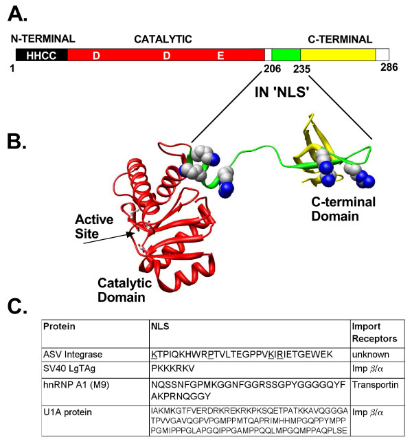
Figure 1.
The ASV IN NLS and three well characterized NLSs. A. Linear map of ASV IN showing the location of NLS sequence. The 286 amino acid IN protein is composed of three domains. The N-terminal, Zn-binding (HHCC) domain (dark) and the central catalytic core domain (red) with the locations of the active site residues (D, D, E) are indicated. The nuclear localization signal, amino acids 206–235 (green), extends from a linker region and into the C-terminal domain (yellow). B. A 3-D structural ribbon model of the catalytic core and C-terminal domains of ASV IN [58] with the with basic residues of the NLS shown in space filling representation. Active site residues in the core domain are shown in ball and stick representation. C. Comparison of the sequences of the ASV IN NLS with three well-characterized NLSs used in the studies reported herein. Residues underlined in the ASV IN NLS have been shown to be required for function.

