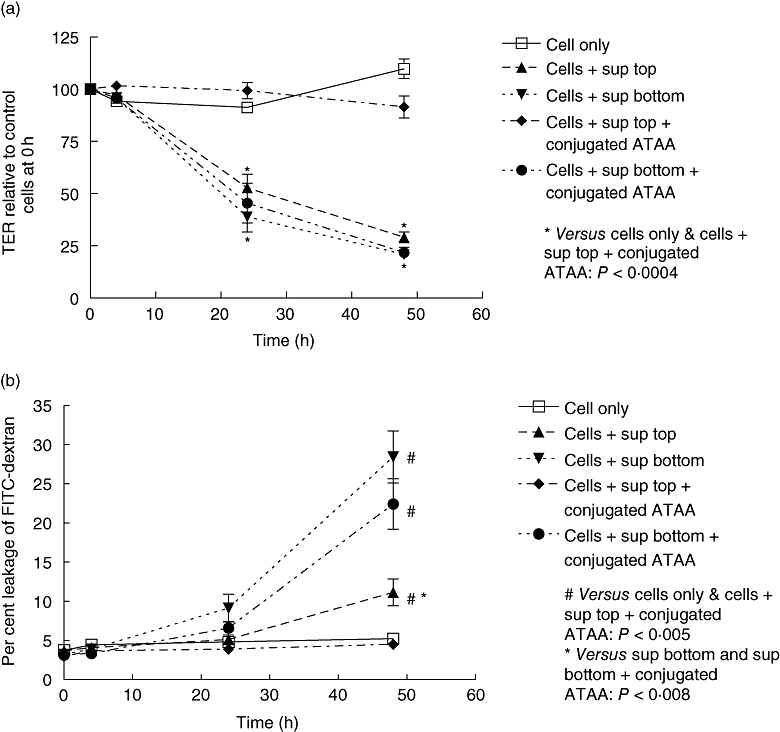
Fig. 3.

Anti-toxin A antibody (ATAA) protects against loss of epithelial barrier function induced by apical, but not basal, application of supernatant sample of VPI 10463 strain of C. difficile. VPI 10463 strain of C. difficile was cultured anaerobically and diluted supernatant samples were pre-incubated with PBS (for 1 h) or sepharose bead-conjugated ATAA, before application to the upper (apical exposure) or lower (basal exposure) chambers of transwells containing confluent monolayers of Caco-2 cells. Transepithelial electrical resistance (TER; a) and permeability to dextran (b) was assessed at 4 h, 24 h and 48 h. The figure represents mean data from four experiments (performed in duplicate). For TER at 24 h and 48 h, cells only ( ) and cells + supernatant + conjugated ATAA to top chamber (
 ) versus the rest: P < 0·0004 (*). The following P values refer to dextran permeability at 48 h; cells only (
 ) and cells + supernatant + conjugated ATAA to top chamber (
 ) versus the rest: P < 0·005 (#). Cells + supernatant to top chamber versus cells + supernatant to bottom chamber, or cells + supernatant + conjugated ATAA to bottom: P < 0·008 (*for both comparisons).
