Figure 2.
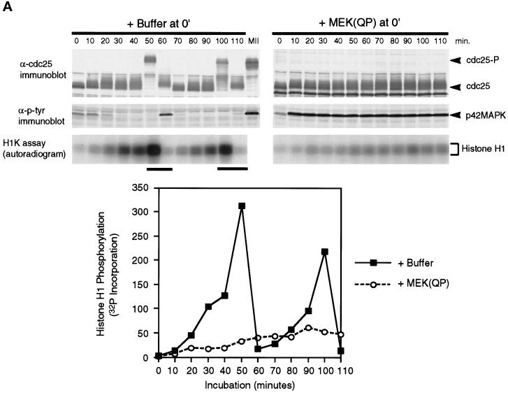
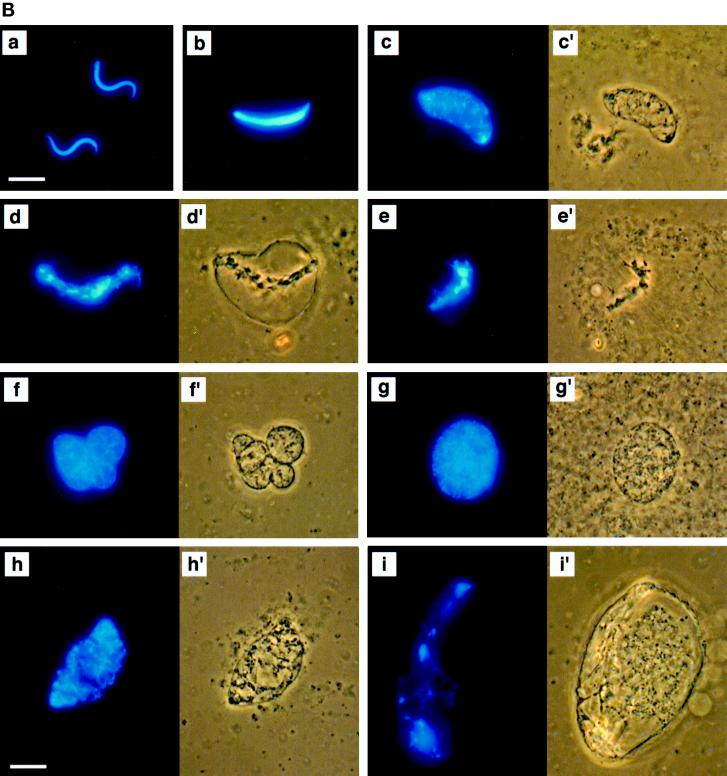
MEK(QP) Activates p42MAPK and inhibits entry into M-phase in cycling Xenopus egg extracts. (A) Cycling egg extracts were prepared and buffer or constitutively active MEK(QP) was added. Reactions were incubated at room temperature and samples were taken for analysis by immunoblotting with Cdc25C antibodies (top) or anti-phosphotyrosine antibodies (middle) and for assay of histone H1 kinase activity (bottom). Bold lines under H1 kinase assays indicate time points when NEBD and chromosome condensation (CC), markers of M-phase, were observed. Bottom, quantitation of H1 kinase activity. (B) Samples were taken for cytology from control cycling extracts or extracts to which MEK(QP) had been added at the start of incubation. Bars, 10 μm. In double adjoining panels, the left image is Hoechst fluorescence and the right image, denoted with ′, is a phase-contrast image. (a–g) Control cycling extracts. (a) Sperm preparation. (b) Sperm nucleus at 10 min. (c and c′) Decondensed nucleus at 20 min. (d and d′) Nucleus just before M-phase at 40 min. (e and e′) NEBD and CC at 50 min. (f and f′) Multiple decondensing nuclei at 70 min. (g and g′) Decondensed nucleus with nuclear envelope at 70. (h and i) Extracts inhibited from entering M-phase by p42MAPK activation in interphase. (h and h′) Decondensed nucleus at 30 min. (i and i′) Nucleus with enlarged intact nuclear envelope at 90 min.


