Figure 3.
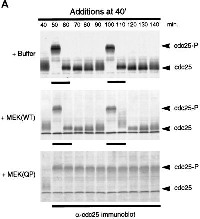
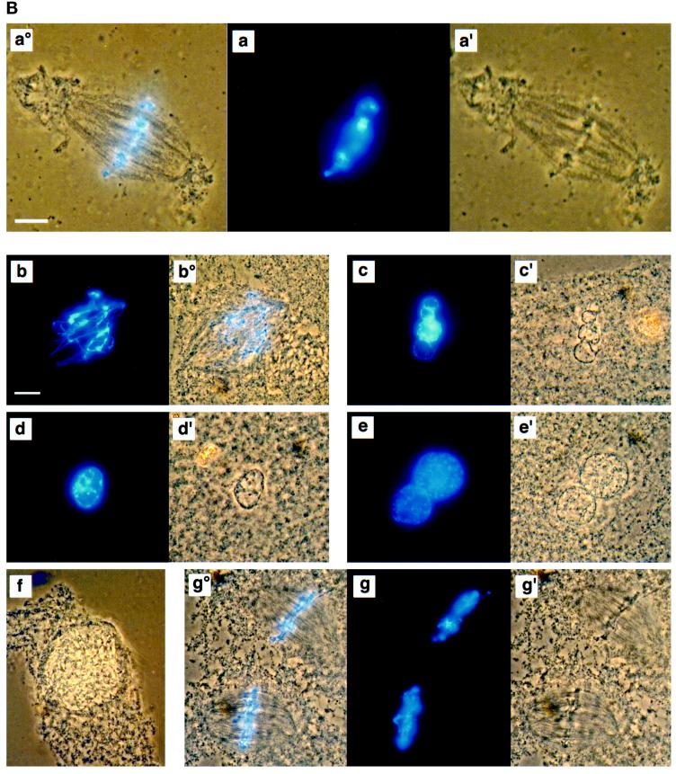
MEK(QP)-induced p42MAPK activation during entry into M-phase in cycling egg extracts leads to a Ca2+-sensitive metaphase arrest. (A) Cycling egg extracts were incubated for 40 min before buffer, MEK(WT), or MEK(QP) was added. Reactions were incubated further and samples were taken for analysis as in Figure 2A. Bold lines under blots indicate time points when M-phase (NEBD and CC) was observed. (B) Samples were taken for cytology as described in Figure 2B after MEK(QP) protein had been added to extracts at 40 min of incubation to activate p42MAPK during entry into M-phase. Extracts arrested in metaphase by MEK(QP) induced activation of p42MAPK during entry into M-phase (sample from A above). (a°, a, and a′) Metaphase spindle at 100 min [60 min after MEK(QP) addition]. Bar, 10 μm. Extracts were arrested in M-phase by the addition of MEK(QP) protein as described above. Thirty minutes later, samples were taken, then Ca2+ (b–f) or double distilled H2O (g) was added, and samples were taken for cytology during further incubation. The samples noted below indicate the duration of incubation after the addition of Ca2+ or double distilled H2O. Bar, 10 μm. Letter alone denotes fluorescence image (Hoechst staining); ′ denotes phase/contrast image; ° denotes fluorescence/phase double exposure. (b and b°) Anaphase at 10 min. (c and c′) Decondensing nuclei at 30 min. (d and d′) Decondensed nucleus at 30 min. (e and e′) Decondensed nuclei at 40 min. (f) Enlarging nucleus at 70 min. (g°, g, and g′) Metaphase spindles at 70 min.


