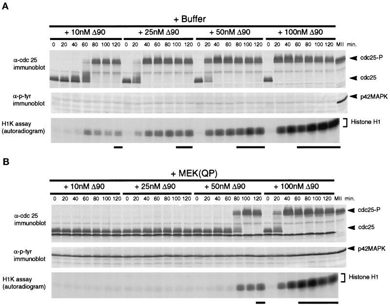
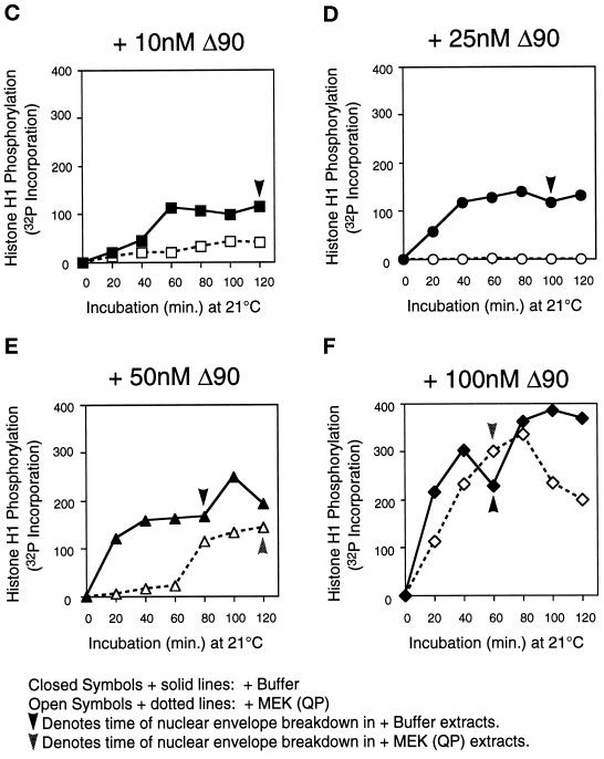
Figure 6.
MEK(QP) inhibits activation of Cdc2 by cyclin BΔ90 in CHX extracts. Extracts were prepared from cycloheximide-treated activated eggs that lack endogenous mitotic cyclins (see MATERIALS AND METHODS). Either buffer or MEK(QP) was added to extracts, after incubation for 20 min. Samples were taken, then increasing concentrations of recombinant cyclin BΔ90 protein were added, and samples were taken as described above. Samples were analyzed by immunoblotting with anti-Cdc25C antibodies (top) and anti-phosphotyrosine antibodies (middle) and assayed for histone H1 kinase activity (bottom). Bold lines below H1 kinase data indicate the time points when NEBD and CC were observed. (A) Sham. Buffer added to reactions, then incubated for 20 min before cyclin BΔ90 addition. (B) MEK(QP) was added to reactions and then incubated for 20 min before cyclin BΔ90 addition. (C–F) Phosphorimager quantitation of histone H1 kinase assay data in A and B above.


