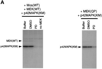
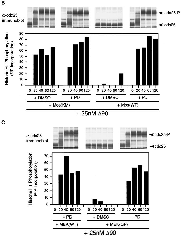
Figure 7.
MEK(QP) and Mos-induced inhibition of Cdc2 activation occurs by specific activation of the p42MAPK signaling pathway. (A) PD inhibits phosphorylation of p42MAPK by MEK(WT) and MEK(QP) in vitro. Left, MEK(WT) was preincubated with buffer, DMSO, or PD (100 μM), and then activity was assayed in vitro by using recombinantXenopus Mos as an activator and recombinant kinase-inactive Xenopus p42MAPK as a substrate. Right, MEK(QP) was preincubated with buffer, DMSO, or PD (100 μM), and then activity was assayed in vitro by using the p42MAPK protein used above as a substrate (see MATERIALS AND METHODS for details). (B) PD prevents the Mos-induced inhibition of Cdc2 activation by cyclin B. DMSO or PD (250 μM) was added to aliquots of CHX extracts on ice, then Mos(WT) or Mos(KM) were added, and reactions were incubated at room temperature for 45 min before the addition of cyclin BΔ90 to 25 nM. Samples were taken for analysis by immunoblotting and H1 kinase assay. (C) PD prevents the MEK(QP)-induced inhibition of Cdc2 activation by cyclin B. DMSO or PD (250 μM) was added to aliquots of CHX extracts on ice, MEK(WT) or MEK(QP) were added, and then reactions were incubated at room temperature for 20 min prior to the addition of cyclin BΔ90 to 25 nM. Samples were taken for analysis by immunoblotting and H1 kinase assay.


