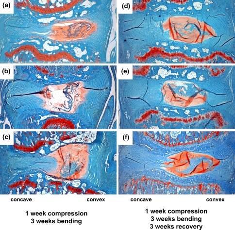
Fig. 6.
Comparison of discs assessed immediately after loading and 3 weeks of bending (a–c) with those assessed after loading, 3 weeks of bending, and 3 weeks of recover (d–f). For all images, the convex side of bending (tissue level tension) is on the right; the concave side of bending (tissue level compression) is on the left. Preservation of anular lamellar structure is clearly evident in all specimens and suggests that no further degenerative changes occurred after the initial 3 weeks of bending during the recovery period

