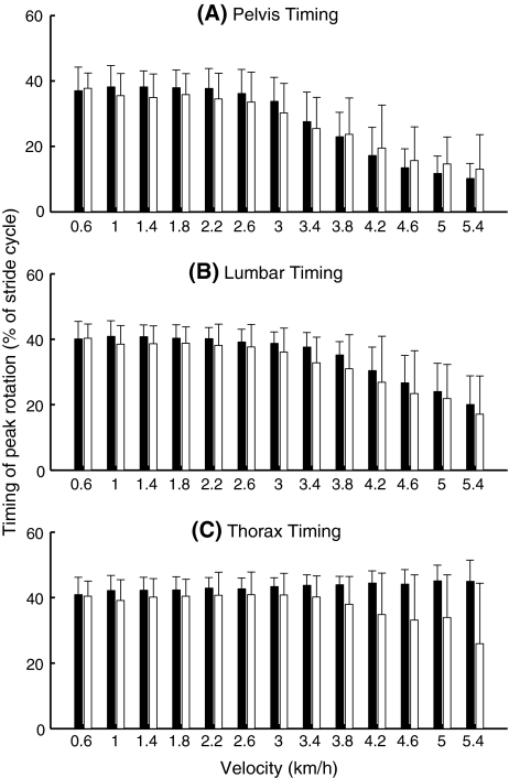
Fig. 4.
Timing of pelvis (a), lumbar segment (b), and thorax (c) peak transverse rotations. Values closer to 0 imply that the rotation was more in synchrony with the upper leg, while values closer to 50 imply that the rotation was more in opposition to the movements of the upper leg. Black bars represent data from the healthy pregnant women, white from the pregnant women with PPP. Error bars represent standard deviations

