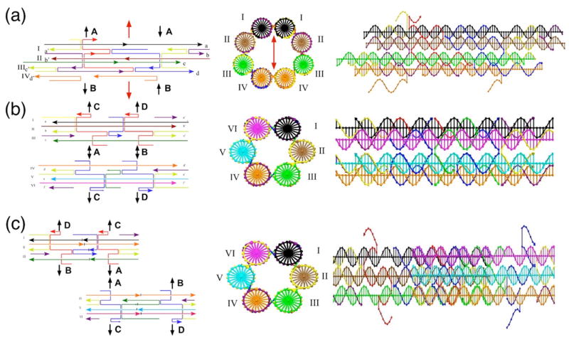
Figure 1.

Schematic Drawings of DNA Half-Tube Molecules. At the left of each panel is a line drawing indicating the structure of the half-tube molecules and the ways in which the molecules connect to each other. These are indicated by letters and arrows that connect to the same letters and arrows. Lateral connections are shown as gaps and unpaired strands. In the center of the panels the cross-sections of the complete tubes are shown schematically, and the helices are labeled with Roman numerals. To the right is a view with the helix and tube axes horizontal. (a) The 4HB half-tube molecule. The central portion shows an elliptical cross-section for the 8HB molecule. The dyad axis relating it to itself is drawn vertically as a red double-headed arrow. Only the 4HB molecule is shown at right. (b) The BTX molecules with similar phasing. (c) The BTX molecules phased half a length apart.
