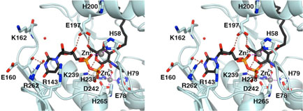
Figure 5.
Model of the LpxC-substrate complex based on the X-ray crystal structures of the TU-514 complex (22) and the UDP complex (Figure 3). Atoms are color coded as follows: C= light cyan O = red, N = blue, P = orange; the substrate carbon atoms appear in black; zinc ions appears as grey spheres; solvent molecules appear as red spheres. Intermolecular interactions observed in the two complexes separately can be achieved by an intact molecule of the substrate, UDP –{3-O-[(R)-3-hydroxymyristoyl]}-N-acetylglucosamine (Figure 2A), bound in the active site.

