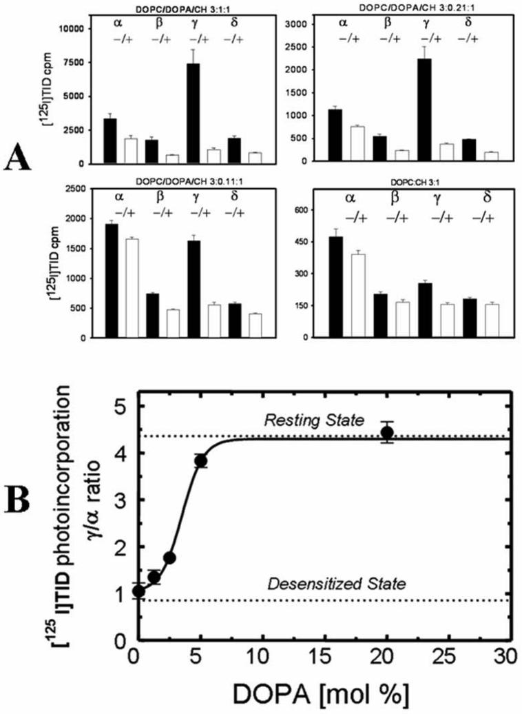
Figure 2. Effect of DOPA levels in DOPC/CH membranes on the photoincorporation of [125I]TID into subunits of the nAChR.
Affinity-purified nAChRs were reconstituted into membranes comprised of DOPC, cholesterol and increasing amounts of DOPA. The molar ratio of each lipid mixture (DOPC/DOPA/CH) was 3:[X]: 1 respectively. Reconstituted membranes were equilibrated for 1 h with [125I]TID (0.4 μM) in the absence (− lanes) and in the presence (+ lanes) of 400 μM Carb, irradiated at 365 nm for 7 min, and polypeptides resolved by SDS-PAGE. A, for each [125I]TID labeling experiment, individual nAChR subunit bands were excised from the dried gel and the amount of [125I]TID photoincorporated into each subunit determined by γ counting (5 min counting time). [125I]TID incorporation into γ' was added to that of the γ-subunit. Shown are bar graphs of the amount of 125I cpm incorporated into each nAChR subunit in the absence or presence of Carb (−/+) and presented as the average of triplicate determinations from a single [125I]TID labeling experiment (error bars indicate the standard error). B, for each [125I]TID labeling experiment, the ratio of the amount of [125I]TID labeling in the γ– and α–subunit in the absence of agonist was calculated. Shown is the relationship between the molar percentage of DOPA in the membrane and the functionality of the nAChR as measured by [125I]TID labeling (γ/α ratio). The γ/α ratio points (●) are means of three different [125I]TID labeling experiments (error bars indicate standard error, see also supporting information). For comparison, the γ/α ratios for nAChRs fully stabilized in the resting and desensitized states are indicated with a dotted line.

