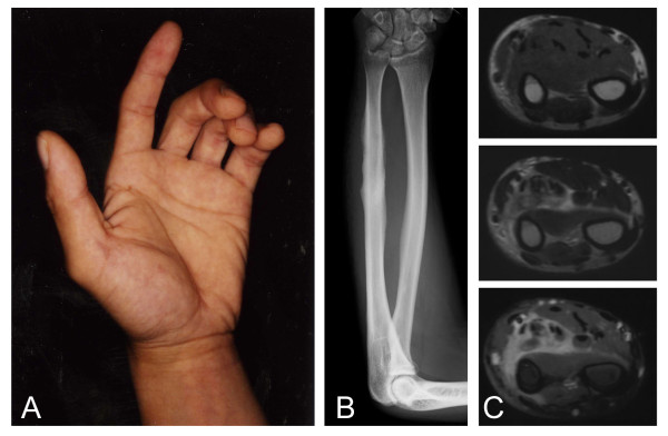
Figure 1.
Epithlioid sarcoma in the forearm. Flexion contracture of the fingers can be seen (A). Plain radiograph shows irregular surface of the ulna (B). MRI of the forearm shows an abnormal lesion with iso-signal intensity on T1-weighted image (top) and high-signal intensity on T2-weighted image (middle) (C). Enhancement with gadolinium can be seen on T1-weighted fat-suppression image (bottom) (C).

