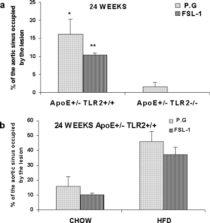
Figure 4. TLR2 activation through FSL-1 demonstrated no significant difference in proximal aortic lesions when compared to P. g in ApoE+/−-TLR2+/+ and ApoE+/−-TLR2−/− mice.
Microscopic cross-sections (10 µm) of the proximal aortic root were stained with Sudan IV and counterstained with hematoxylin to reveal lipid deposition, which was quantified by digital morphometry for samples from ApoE+/−-TLR2+/+ and ApoE+/−-TLR2−/− mice. (4A): percentage of total lumen of the proximal aorta occupied by lesions after 24 weeks of treatment in ApoE+/−-TLR2+/+ and ApoE+/−-TLR2−/− mice maintained on a standard chow diet and injected weekly with P. g or FSL-1. Values represent means±SD; *p<0.05 for differences between mice injected with P. g; **p<0.05 for differences between mice injected with FSL-1. No lesions were detected in ApoE+/−-TLR2−/− mice irrespective of the treatment. (4B): percentage of total lumen of the proximal aorta occupied by lesions in ApoE+/−-TLR2+/+ mice maintained on a chow diet or a high fat diet after 24 weeks of injections with P. g or FSL-1. Values represent means±SD.

