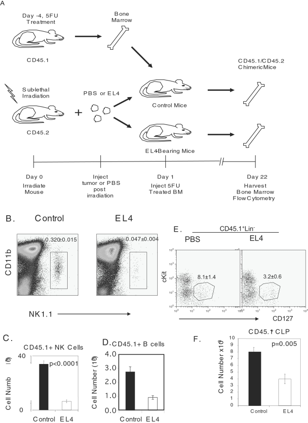
Figure 6. Decrease in CLPs, NKPS and Block in NKP progression.
B6 mice were sublethally irradiated with 500 Rad prior to EL4 or PBS injection. Twenty-four hours later 5FU treated bone marrow from congenic CD45.1 mice was injected into control or EL4-bearing mice. Three weeks later bone marrow was harvested to evaluate NK cell progenitors. Donor NK cells were identified by an anti-CD45.1. A. Diagram of experiments. B. FACs profile of CD45.1+ NK1.1+ cells. Data shown are from gated CD45.1+CD3− cells. The numbers shown in the panels are means and SEM, involving a total of 5 mice per group. C. Absolute number of CD45.1+ NK cells. D. Absolute number of donor B cells. E. FACS profile of donor CLP, characterized as CD45.1+Lin−CD127+cKitint. Data are representative of 5 mice per group. F. Absolute number of CD45.1+ and CD45.1− CLP. Data shown are means and SEM, involving a total of 5 mice per group.

