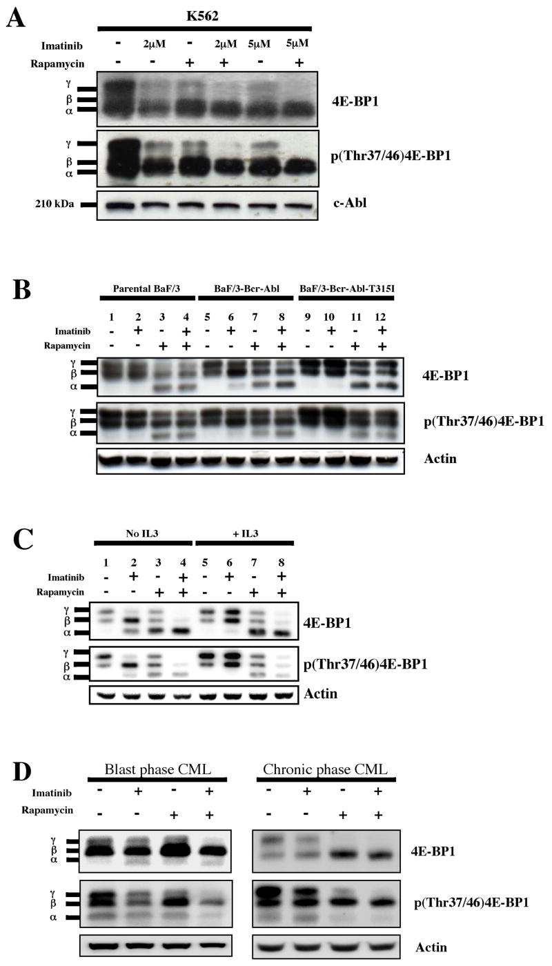
Figure 1.
Bcr-Abl and mTORC1 contribute to 4E-BP1 phosphorylation in CML cells. (a) K562 cells were treated for 24 hrs with diluent (DMSO) alone, imatinib (2 and 5 μM), rapamycin (10 ng/ml), or both. Western analysis was performed on whole cell lysates using antibodies to total 4E-BP1, phospho(Thr37/46)4E-BP1, and c-Abl as a loading control. The antibody to total 4E-BP1 recognizes all forms of 4E-BP1. The α, β, and γ bands correspond to increasingly phosphorylated forms of 4E-BP1. (b) Parental Ba/F3, Ba/F3-Bcr-Abl and Ba/F3-Bcr-Abl-T315I cells were treated for 4 hrs with diluent alone, imatinib (2 μM), rapamycin (10 ng/ml), or both. Immunoblot was performed as described above. Anti-actin was used as a loading control. (c) Ba/F3-Bcr-Abl cells were treated with or without inhibitors for 4 hrs in the presence or absence of IL3, and immunoblot performed. For IL3-treated cells, 10 ng/ml of the cytokine was added 12–16 hrs before the addition of inhibitors. (d) Primary CML cells from patients in blast and chronic phase CML obtained at the time of presentation were grown in serum-free media and GF’s. Cells were incubated with the indicated inhibitors for 24 hrs, and immunoblot analysis performed on cell lysates. Antibody to actin was used as a loading control.

