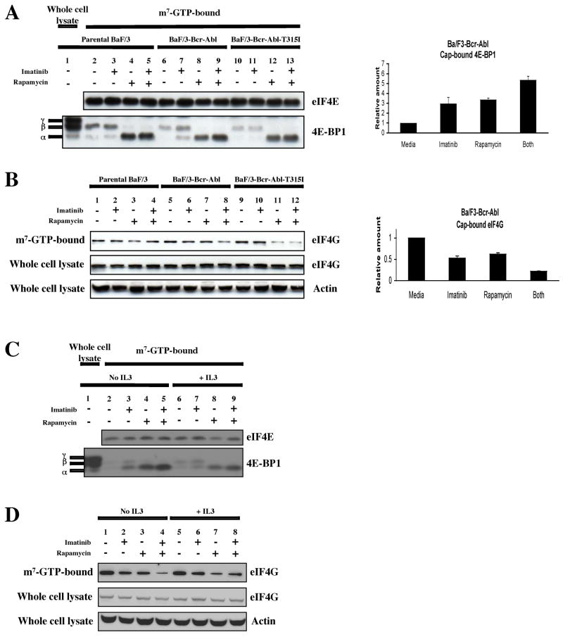
Figure 3.
Bcr-Abl induces the formation of eIF4F in Ba/F3-Bcr-Abl, but not parental Ba/F3 or Ba/F3-Bcr-Abl-T315I cells. (a) Left-hand panel: Bcr-Abl and mTOR decrease the amount cap-bound 4E-BP1 in Ba/F3-Bcr-Abl cells. Parental Ba/F3, Ba/F3-Bcr-Abl and Ba/F3-Bcr-Abl-T315I cells were treated for 4 hrs with diluent alone, imatinib (2 μM), rapamycin (10 ng/ml), or both. Right-hand panel: Quantitation of cap-bound 4E-BP1 in Ba/F3-Bcr-Abl cells. Bars indicate standard errors calculated from three independent experiments. (b) Left-hand panel: Bcr-Abl and mTOR promote the binding of eIF4G to cap-bound eIF4E. The same blots described in (a) were probed with antibody recognizing eIF4G. In addition, immunoblot of whole cell lysates were probed to assess total levels of eIF4G. Equal loading was assessed by an anti-actin antibody. Right-hand panel: Quantitation of cap-bound eIF4G in Ba/F3-Bcr-Abl cells. Bars indicate standard errors calculated from three independent experiments. (c) and (d) Addition of IL3 partially abrogates the inhibitory effect of imatinib on eIF4F formation in Ba/F3-Bcr-Abl cells. Ba/F3-Bcr-Abl cells were treated and analyzed as in (a) and (b), but in the presence and absence of IL3. For IL3-treated cells, 10 ng/ml of the cytokine was added 12–16 hrs before the addition of inhibitors.

