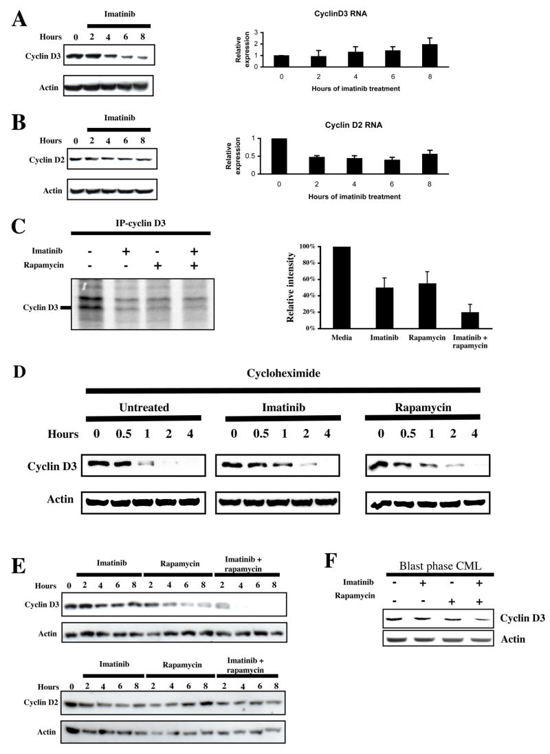
Figure 5.
Bcr-Abl and mTORC1 regulate cyclin D3, but not cyclin D2, translation. (a) Ba/F3-Bcr-Abl cells were incubated with 2 μM imatinib, and sampled every 2 hrs for 8 hrs. Western analysis of cell lysates and Q-PCR of total RNA was performed to assess cyclin D3 protein and mRNA levels. Bars indicate standard errors calculated from three independent experiments. (b) Cyclin D2 protein and mRNA levels were also assessed as in A. Bars indicate standard errors calculated from three independent experiments. (c) Ba/F3-Bcr-Abl cells were incubated in serum- and methionine-free media for 3 hrs, following which they were metabolically labeled with S35-methionine, and treated with or without imatinib, rapamycin, or both for 3 hrs. Cyclin D3 was immunoprecipitated, separated by SDS-PAGE, and S35-labelled cyclin D3 visualized by autoradiography. The intensity of the band corresponding to cyclin D3 was averaged from three independent experiments and plotted as a bar graph. Bars indicate standard errors. (d) Ba/F-Bcr-Abl cells were pretreated with diluent, 2 μM imatinib, or 10 ng/ml rapamycin for one hour to ensure kinase inhibition, following which 20 μg/ml cycloheximide was added. Cells were then harvested and lysed at the indicated times. Cyclin D3 protein levels were assessed by immunoblot. Actin levels were used as a loading control. (e) Ba/F-Bcr-Abl cells were treated with 2 μM imatinib, 10 ng/ml rapamycin, or both over 8 hrs. Cyclin D2 and cyclin D3 protein levels were assessed by immunoblot. (f) Primary CML cells were grown in GF-supplemented serum-free media, and treated with or without imatinib (2μM), rapamycin (10 ng/ml), or both for 24 hrs. Immunoblot for cyclin D3 and actin was performed on lysates obtained from these cells.

