FIG. 5.
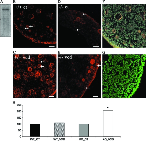
Effect of VCD on mEH protein. (A) Representative Western blotting of adult B6C3F1 mouse ovary protein incubated with anti-mEH antibody. Ovaries from PND4 CYP2E1+/+ (B, C) and CYP2E1−/− (D, E) mice were incubated with control medium (B, D) or 5μM VCD (C, E) for 15 days. Following culture, ovaries were processed for immunofluoresence and confocal microscopy as described in the Materials and Methods section. All images were captured with a ×40 objective lens. Red staining = Cy5-labeled anti-mEH antibody. Green staining = YoYo1 genomic DNA. (F) Merged genomic stain and mEH protein stain. (G) Immunonegative stain (Cy5 and YoYo1 without anti-mEH antibody). (H) Quantification of mEH staining intensity, relative to controls, in small preantral follicles. Broken arrows indicate primordial follicles. Solid arrows indicate small primary follicles. Scale bar = 25μM.

