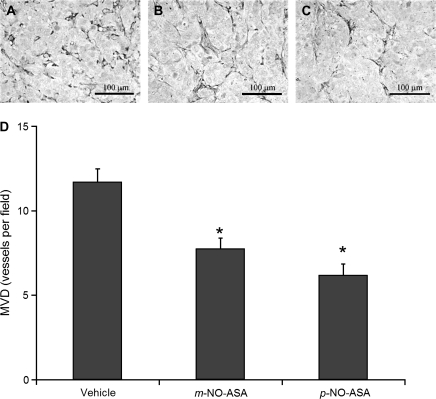
Fig. 3.
NO-ASA reduces MVD in HT-29 xenografts. Representative sections of tumors injected with (A) vehicle, (B) m-NO-ASA or (C) p-NO-ASA that show endothelial cell staining using the anti-PECAM-1 antibody. There were 12 mice per group. The number of microvessels within a given area was determined as in Materials and Methods and their mean ± SEM values are depicted in the graph below. *Statistically significant difference compared with the vehicle control group. Original magnification ×200. The bar represents 100 μm.

