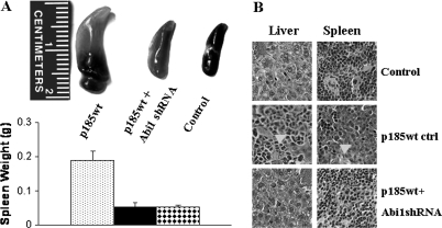
Fig. 5.
Pathology analysis of the mice injected with Ba/F3 cells, control p185wt cells and p185wt cells transduced with Abi1 shRNA. (A) Spleen weight of mice injected with Ba/F3 cells (control) and the p185wt cells expressing with (p185wt + Abi1 shRNA) or without (p185wt) Abi1 shRNA. (B) Histology of spleens and livers from the mice receiving Ba/F3 cells (control) and the p185wt cells expressing with (p185wt + Abi1 shRNA) or without (p185wt ctrl) Abi1 shRNA. Arrowheads indicate the massively expanded p185wt cells.

