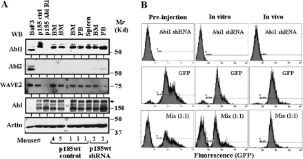
Fig. 6.
Abi1 knockdown impeded in vivo competitive expansion of p185wt cells. (A) Expression of Abi1 in Ba/F3 cells, p185wt cells transduced with non-silencing shRNA (p185wt control) or Abi1 shRNA (p185wt Abi1 Ri), as well as the cells rescued from bone marrow (BM), peripheral blood (PB) and spleen of the diseased mice injected with p185wt cells transduced with either non-silencing shRNA or Abi1 shRNA. The 100 μg total proteins from these cells were separated by sodium dodecyl sulfate–polyacrylamide gel electrophoresis, transferred to nitrocellulose membrane and western blotted (WB) with indicated antibodies. Molecular markers are indicated. The arrow indicates p185Bcr-Abl. (B) In vivo competitive expansion of p185wt GFP cells and p185wt Abi1 shRNA cells. The p185wt cells were transduced with MSCV-based retroviruses expressing either GFP or Abi1 shRNA. The GFP-positive p185wt cells were sorted by fluorescence-activated cell sorting to a purity of >95%. The GFP-positive cells then were mixed with p185wt cells expressing Abi1 shRNA at a ratio of 1:1. The mixed cells were either cultured in vitro for 5 weeks or injected into Balb/c mice (1 × 10⁶ cells per mouse) through tail vein. The cells derived from p185wt cells were rescued from bone marrow and peripheral blood (data not shown) of the diseased mice by selection with puromycin. The rescued cells and the cells cultured in vitro were subjected to flow cytometry analysis.

