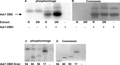
Figure 1. Phosphorylation of Adr1 DBD by cell extracts.
A. In vitro kinase assay with purified, recombinant Adr1 DBD (amino acids 17–160 of Adr1), [γ-32P]-ATP and cell extracts from TYY201. R, cells were grown in repressing medium; DR, cells were derepressed in 0.05% glucose for 3 hours. Because of variability in the extracts, quantitative comparisons cannot be made between the repressed and derepressed lanes. B. Coomassie stain of the gel in A. C. In vitro kinase assay as in A, but with N-terminally truncated versions of the Adr1 DBD. Numbers indicate the amino acid in wild-type Adr1 that is the N-terminus of the truncated version. D. Coomassie stain of the gel in C.

