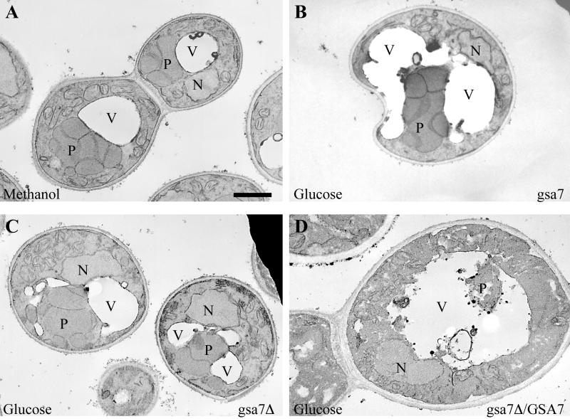
Figure 2.
Morphology of gsa7 and gsa7Δ cells during glucose adaptation. gsa7Δ cells were grown in methanol (A) and gsa7, gsa7Δ, and gsa7Δ cells transformed with the normal GSA7 gene (gsa7Δ/GSA7) were grown in methanol induction medium then adapted to glucose for 3 h (B–D). Cells were harvested, fixed in potassium permanganate, and prepared for electron microscopy as described in MATERIALS AND METHODS. N, nucleus; V, vacuole; P, peroxisome. Bar, 0.5 μm.

