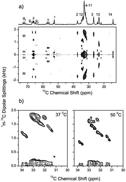
Fig. 3.
(a) A 2D PDLF spectrum of DMPC/DHPC bicelles at 37°C. 64 scans were accumulated for each of 200 points in the t1 dimension with an increment time of 384 μs. The contact time for the CP transfer was set to 3.0 ms. Proton RF field during the t1 evolution corresponded to 31 kHz. A 1D 13C chemical shift spectrum is shown at the top with assignments of peaks to individual carbons of the DMPC molecule. (b) Part of the PDLF spectrum in DMPC/DHPC bicelles at 50°C demonstrating the increased dipolar resolution in the crowded spectra range between 31 and 34 ppm. Corresponding part of the PDLF spectrum obtained at 37°C is also shown for comparison.

