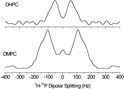
Fig. 7.
Cross sections from the 2D 31P-1H PDLF spectrum at the chemical shift of the 31P resonance in DMPC and DHPC molecules. 64 transients were accumulated for each of 32 points in the t1 dimension with an increment time of 2300 μs.
The location of a desipramine molecule relative to a DMPC lipid in bicelles. A change in the head group conformation of DMPC is also indicated. The relevant hydrogen atoms are included in the figure, while all other hydrogen atoms are not represented.

