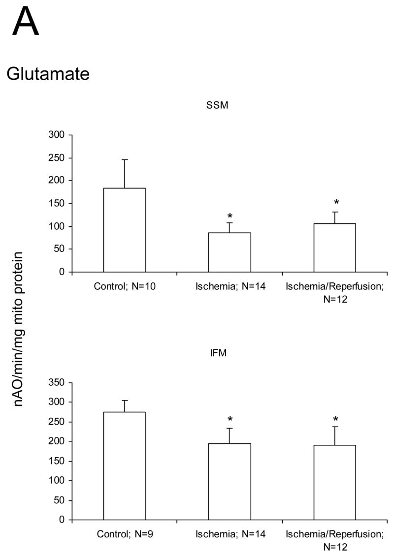
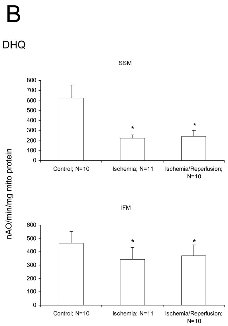
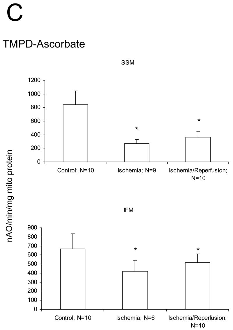
Figure 1.
Ischemia markedly decreases the maximal ADP-stimulated rate of oxidative phosphorylation in both subsarcolemmal (SSM) and interfibrillar (IFM) mitochondria obtained from the 24 month old Fischer 344 rat heart. Reperfusion does not result in additional decreases in the rate of oxidative phosphorylation. Decreases are observed with glutamate (A), duroquinol (B), and TMPD-ascorbate (C) as substrates. The decrease in the rate of respiration are similar in the mitochondria isolated both following 25′ ischemia and following 25′ ischemia plus 30′ reperfusion. (Mean ± SD; * p<0.05; p=NS Ischemia vs. Ischemia-Reperfusion.)


