Figure 1.
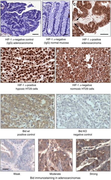
Experimental controls for immunohistochemistry. (A) Substitution of the primary antibody with mouse IgG1 at the same dilution was used as a negative control in all experiments. (B) Normal large bowel mucosa was an additional IgG1-negative control. (C) Representative immunostaining for HIF-1α demonstrating immunopositivity in close proximity to blood vessels (arrows). Both nuclear and cytoplasmic staining is seen. (D and E) Formalin-fixed and paraffin-embedded pellets of HT-29 human colorectal cancer cells previously cultured for 16 h in normoxic or hypoxic conditions (1% O2) and stained for HIF-1α. Only nuclear staining, as seen in hypoxic cells, was considered positive; we considered the diffuse cytoplasmic staining in normoxic cells as nonspecific, reasoning that HIF-1 is not transcriptionally active in cytosol. (F and G) Formalin-fixed and paraffin-embedded pellets of Bid knockout and wild-type mouse embryonic fibroblasts (kindly provided by SJ Korsmeyer, Harvard Medical School, Boston, MA, USA) were used as positive and negative controls for Bid. Pellets were processed identical to that for human tumour specimens. (H–J) Bid immunostaining in human adenocarcinomas with representative sections of weak (score 1), moderate (score 2), and strong (score 3) staining. The magnification bar for all sections is 50 μm.

