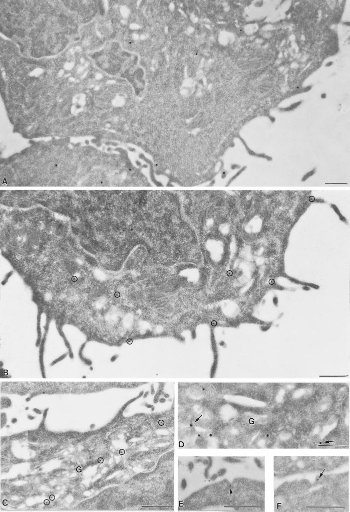
Figure 7.
Immunoelectron microscopy of PLCγ2. anti-PLCγ2 was localized in thin sections of LR White-embedded resting (A) and antigen-activated (B-F) RBL-2H3 cells by labeling with anti-PLCγ2 followed by 15 nm Protein A-gold with a rabbit anti-mouse bridge (B,C,E,F) or with 30 nm anti-mouse IgG-colloidal gold particles (A,D). 15 nm gold particles in B,C are circled for emphasis. Gold particles marking PLCγ2 associate with the plasma membrane and also prominently label vesicles in the Golgi region (G) in panels C,D. Occasional gold particles label Golgi stacks (arrows, D). Gold labeling was also found in association with coated pits (E,F). Micrographs are representative of results in 3 different experiments. Bar = 0.5 μm.

