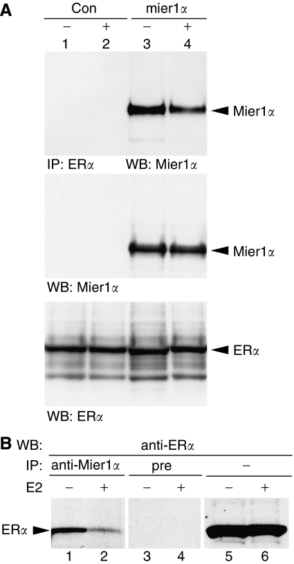
Figure 1.
MI-ER1α interacts with ERα in vivo. (A) HEK293 cells were transfected with pcDNA3-herα and either pCS3+MT-mier1α (lanes 3 and 4) or control empty vector (lanes 1 and 2) and treated with vehicle (lanes1 and 3) or 10 nM E2 (lanes 2 and 4) for 3 h before extraction. Extracts were subjected to immunoprecipitation with anti-ERα (top panel) or loaded directly onto the gel (middle and bottom panels). Western blots were stained for MI-ER1α (top and middle panels) or ERα (bottom panel). (B) Extracts from MCF-7 cells treated with vehicle (lanes 1, 3 and 5) or 10 nM E2 (lanes 2, 4 and 6) were subjected to IP with anti-mier1α (lanes 1 and 2), pre-immune (lanes 3 and 4) or loaded directly onto the gel (lanes 5 and 6); Western blotting was performed with anti-ERα.

