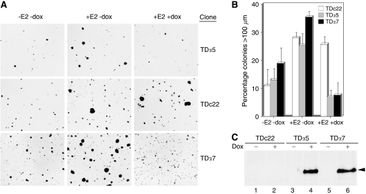
Figure 2.
Overexpression of MI-ER1α reduces anchorage-independent growth of T47D breast carcinoma cells. Control (TDc22)- or MI-ER1α (TDα5 and TDα7)-expressing Tet-On T47D clones were cultured in 0.35% agarose, in the presence or absence of 2 μg ml−1 dox and in the presence or absence of 10 nM E2, as described in the Materials and methods. Colonies were stained with crystal violet, and colony size was measured using an ocular micrometre. A minimum of six fields from each plate was analysed; the number of colonies larger than 100 μm in size, expressed as a percentage of the total number of colonies, was recorded for each treatment. (A) Representative fields for each treatment combination for each clone are shown. (B) Histogram showing the average values and error bars for three independent experiments, performed in duplicate. (C) Western blot analysis to verify dox-specific induction of MI-ER1α expression. A representative blot of extracts from TDc22, TDα5 and TDα7 cells, cultured in the absence (lanes 1, 3 and 5) or presence (lanes 2, 4 and 6) of 2 μg ml−1 dox, is shown. The position of MI-ER1α is indicated.

