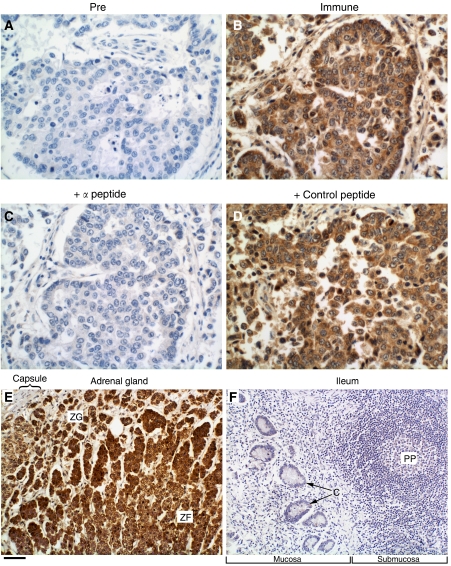
Figure 3.
Immunohistochemical staining with anti-MI-ER1α. Human breast tumour sections were stained with pre-immune IgG (A), anti-mier1α IgG (B) or anti-mier1α IgG that had been pre-incubated with the α-specific peptide (C) or a control peptide (D). Note that only the peptide used to generate the antibody blocks staining. Sections of normal human adrenal gland (E) and normal human small intestine (F) stained with anti-mier1α IgG served as positive and negative tissue controls, respectively. Panel E shows a portion of the adrenal cortex that includes part of the zona glomerulosa (ZG) and zona fasciculata (ZF), whereas Panel F shows a section through the ileum that includes lymphoid tissue (Peyer's patch (PP)) and crypts of Lieberkühn (C). Scale bar=50 μm for A–D and 100 μm for E and F.

