Figure 7.
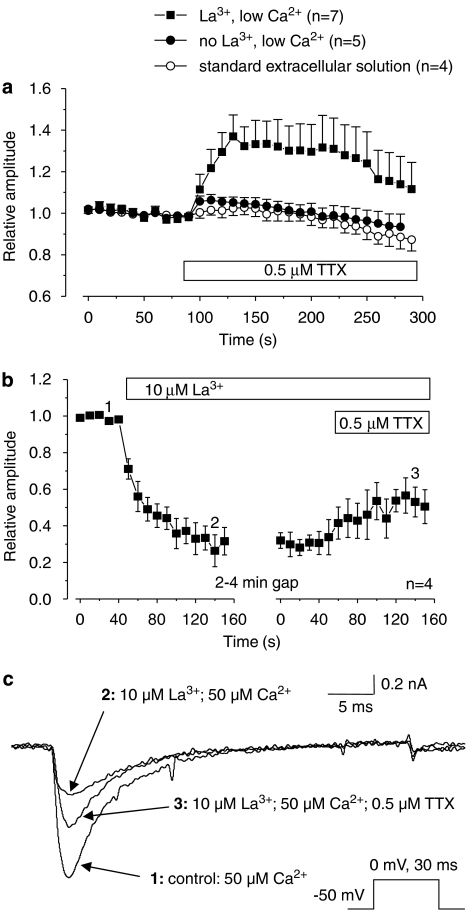
The involvement of extracellular La3+ in the enhancing effect of TTX on isolated TTXR currents. (a) The graph shows relative amplitudes of isolated TTXR current in modified extracellular solutions, with the bar showing the application of 0.5 μM TTX. TTXR currents were evoked as described in Figure 2. Reducing extracellular Ca2+ did not alter the enhancing effect of TTX on TTXR current. Reduction of Ca2+ plus removal of La3+ substantially reduced any current increase by TTX, suggesting that the effect is dependent on the presence of extracellular La3+. In control experiments, recorded in standard extracellular solution, application of TTX-free extracellular solution to the cells had no effect on TTXR current amplitude. (b) The graph shows relative amplitudes of isolated TTXR current recorded in a low extracellular Ca2+ solution (50 μM) in the absence of extracellular La3+ and TTX. TTXR currents were evoked as described in Figure 2. The external application of 10 μM La3+ caused a sustained reduction in TTXR current amplitude of 60–70%. Subsequent addition of 0.5 μM TTX produced a partial reversal of this inhibition. The numbered data points on the graph correspond to the time points of the example traces shown in panel (c). (c) Example traces from data shown in panel b demonstrating the inhibition of TTXR current amplitude by 10μM La3+, and the ability of 0.5 μM TTX to reverse this inhibition by La3+.

