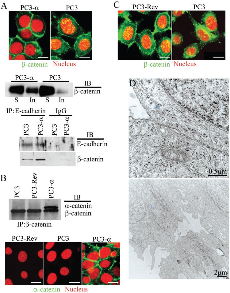
Figure 2. Nuclear/cytoplasmic localization of β-catenin in α-catenin positive and negative PC3 cells.
A, Top panel: Immunofluorescence staining of β-catenin (green) in PC3 and PC3-α cells. Propidium iodide (red) marks the nucleus. β-catenin localized to the nucleus appears in yellow. Bar represents 15 μm. Middle panel: Immunoblot analysis of β-catenin in detergent-soluble (S) and insoluble (In) fractions. Bottom panel: β-catenin co-immunoprecipitating with E-cadherin. IgG was used as a negative control. B, Top panel: Co-immunoprecipitation of α-catenin and β-catenin from PC3, PC3-Rev, and PC3-α cells. The blots were probed with both α- and β-catenin antibodies simultaneously. Bottom panel: Immunofluorescence staining of α-catenin (green) in PC3, PC3-Rev and PC3-α cells. Bar represents 15 μm. C, Immunofluorescence staining of β-catenin in PC3, and PC3-Rev cells. PC3-Rev cells show cytoplasmic (green) and nuclear (yellow) staining of β-catenin comparable to PC3 cells. Propidium iodide (red) marks the nucleus. Bar represents 15 μm. D, TEM of PC3-Rev cells.

