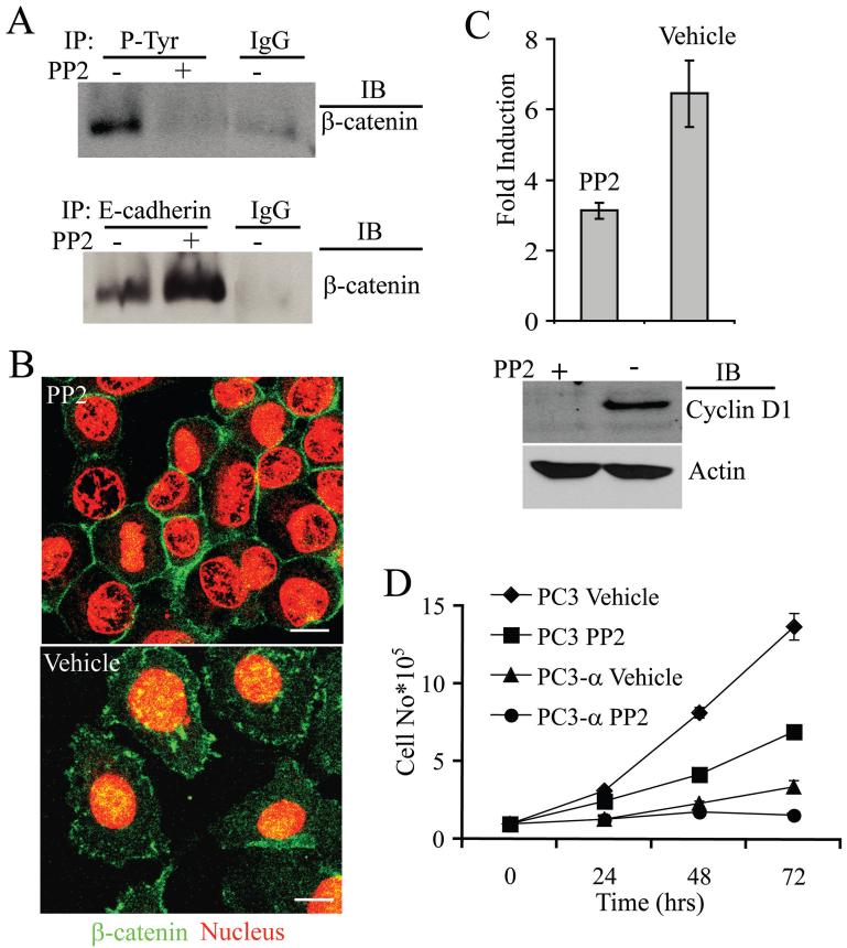
Figure 5. Src regulates β-catenin localization and transcriptional activity in PC3 cells.
A, β-Catenin immunoblot of immunoprecipitates of tyrosine phosphorylated proteins from PC3 cells (upper blot) and of immunoprecipitates of E-cadherin (lower blot). Treatment with PP2 increased the amount of β-catenin associated with E-cadherin. IgG was used as control. Blots represent data from three independent experiments. B, Immunofluorescence staining of β-catenin (green) of PP2 and vehicle-treated PC3 cells. Propidium iodide stains the nucleus (red). Bar represents 15 μm. C, Src inhibition affects β-catenin mediated transcription in PC3 cells. Top Panel-β-catenin/TCF/LEF mediated transcriptional activity. Bars represent standard error of the means of two independent experiments performed in triplicate. Bottom panel: Immunoblot analysis of cyclin D1 levels in PC3 cells treated with either PP2 or vehicle (DMSO) for 48 h. Actin was used as a loading control. Blot represents two independent experiments. D, Effect of Src inhibition on growth of PC3 and PC3-α cells. Twenty-four hours after plating, PP2 or vehicle (DMSO) was added to the media and incubated for an additional 48 h before counting of the cells. Bars represent standard error of the means of two independent experiments done in triplicate.

