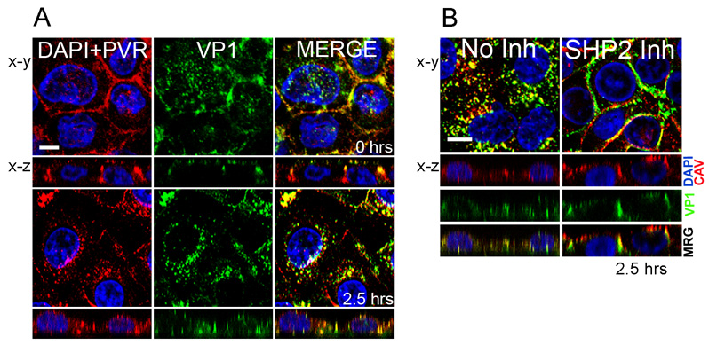
Figure 2. Poliovirus entry in brain microvascular endothelial cells.
(A) Virus enters the cell and moves to a perinuclear compartment. At 0 hrs, virus stained with anti-VP1 antibody (green) is seen at the cell surface most evident in x-z images. Poliovirus receptor (PVR, red) is also at the surface, largely concentrated at intracellular contacts. Cell nuclei are stained with DAPI (blue). By 2.5 hours post-infection, both virus and PVR have moved from the surface to a perinuclear compartment. (B) SHP-2 is required for virus entry. In control cells, virus (VP1, green) is seen within the cell, where it colocalizes with caveolin (CAV, red). In cells treated with an inhibitor of SHP-2 phosphatase, virus remains at the cell surface, largely at intercellular contacts. (Figure adapted from reference 17).

