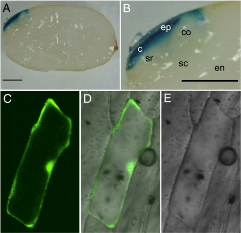
Fig. 6.
GUS expression and subcellular localization. (A) GUS expression under the control of the qLTG3–1 gene promoter. Transgenic plants expressing GUS under the control of the 2-kb upstream region of qLTG3–1 were stained for GUS activity. The seeds were incubated for 1 day. The bars are 1 mm. (B) Close up view of the embryo in (A). c, coleorhiza; co, coleoptile; en, endosperm; ep, epiblast; sc, scutellum; sr, seminal root. (C–E) Subcellular localization of GFP in onion epidermal cells expressing a qLTG3–1-GFP fusion as shown by confocal microscopy.

