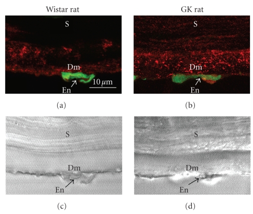
Figure 4.
Immunofluorescence localization of type VIII collagen (red color) in Descemet's membrane of the cornea from 62-week-old rats (a), (b) and Nomarsky microscopic images of the same optical fields (c), (d). (a), (c) Wistar rat. (b), (d) GK rat. Green color (SYBR-Green I) indicates the nuclei of endothelial cells. Dm: Descemet's membrane; En: endothelial cell; S: stroma. Scale bar: 10 μm.

