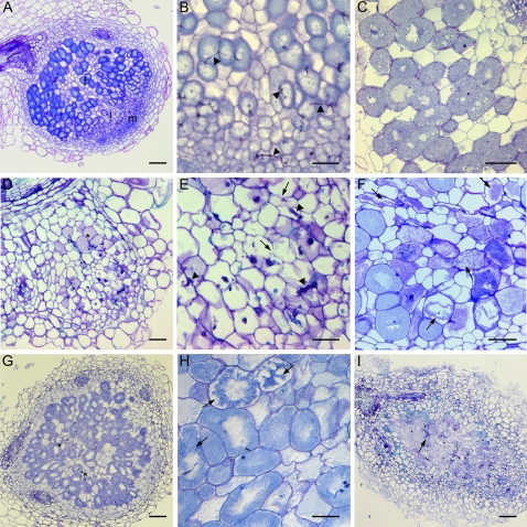
Figure 8.
Bright-field microscopy on 35S:SINAT5DN and control M. truncatula nodules. A, Young wild-type nodule containing a nodule meristem (m), infection zone (i), and fixation (f) zone. B, Detail of infection zone of young control nodule. Arrowheads indicate ITs. C, Wild-type fixation zone (25 dpi) with infected cells filled with symbiosomes. D, Medial section through a young 35S:SINAT5DN nodule with a disordered central tissue and few infected cells (asterisks). E, Detail of infected region in D. Thicker ITs (arrowheads) appear and cells show signs of degradation (arrows). F, Senescent nodule tissue of a wild-type M. truncatula nodule (61 dpi). Arrows mark cells with senescent features. G, Medial section through a 35S:SINAT5DN nodule more developed than the 35S:SINAT5DN nodule in D. Asterisks show senescing regions. H, Detail of G with arrows indicating cells with signs of senescence. I, Cylindrical 35S:SINAT5DN nodule demonstrating early senescence of the nodule central tissue. Bars = 100 μm (B–F and H), 200 μm (A and G), and 400 μm (I).

