Figure 9.
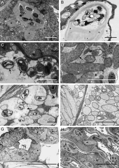
Transmission electron microscopy of 35S:SINAT5DN and control M. truncatula young and mature nodules. A and B, Cross sections through an IT of a 35S:SINAT5DN nodule (A), with fewer bacteria and a denser matrix than a wild-type IT (B). Asterisks indicate bulge-like structures. C, Deformed symbiosomes (arrows) in 35S:SINAT5DN-infected cells. D, Wild-type symbiosomes. The symbiosome membrane tightly encloses a single bacteroid. Arrow marks symbiosome space. E, Hampered 35S:SINAT5DN symbiosome development leading to large symbiosomes containing several degrading bacteroids. F, Section through infected cells of a control nodule. Each bacteroid is surrounded by a symbiosome membrane. G, Cell wall appositions (asterisks) as a sign of plant cell defense in 35S:SINAT5DN nodule tissue. H, Loss of plant cell integrity in the 35S:SINAT5DN nodules displaying the initiation of senescence. Bars = 1 μm (A–G) and 2 μm (H).

