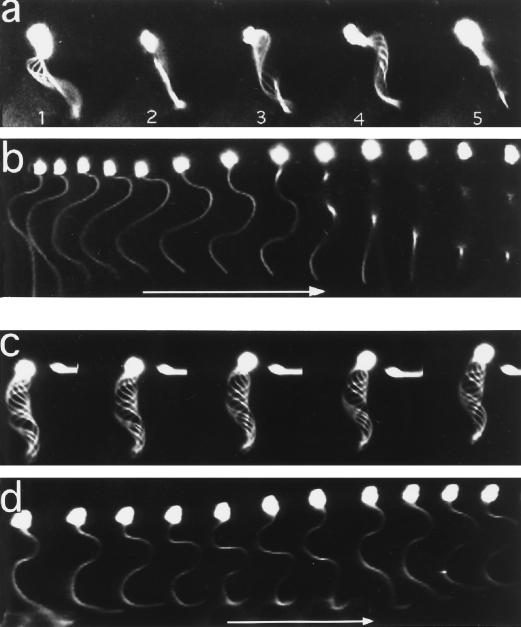
Figure 1.
Video images of demembranated/reactivated sperm models. Sperm models were incubated with (c and d) or without (a and b) D-316 and analyzed by dark-field microscopy using 300-Hz stroboscopic illumination and a translation of the microscope stage (b and d). (a and b) control sperm models without antibody at 171 and 178 s after reactivation; (c and d) Sperm models with D-316 (17 μg/ml) at 176 and 179 s; (a and c) overlapping images; (b and d) individual images taken 3 ms apart. Arrow, 15 μm.

