Figure 4.
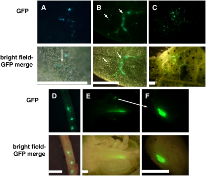
CLCrV:GFP expression is limited to vascular tissue and extends to the ovule integument. Top section shows fluorescence from the smRSGFP-expressing CLCrV vector and the bottom section shows the merged brightfield-GFP photo. Plants were grown at 25°C/23°C and similar, mock-inoculated tissues were negative for GFP-like fluorescence (data not shown) unless mentioned. A, Cotyledon 9 dpi. Arrow indicates gold particles. B, Cotyledon tissue showing viral DNA accumulation along some of the veins, indicating the beginning of systemic movement. Arrows show glandular trichomes that were also autofluorescent in control leaves of the same age. Otherwise, controls showed no fluorescence. C, Older leaf showing GFP-expressing cells associated with vascular tissue. Only some of the veins showed GFP fluorescence, demonstrating that veins are not inherently autofluorescent. D, Root tissue with three regions of CLCrV:GFP DNA accumulation. E and F, Longitudinal section of a 3-DPA ovary showing green fluorescence along part of the central column and in one of the ovules, which is magnified in F. The ovule showed one area with strong GFP fluorescence. Bars = 2 mm.

