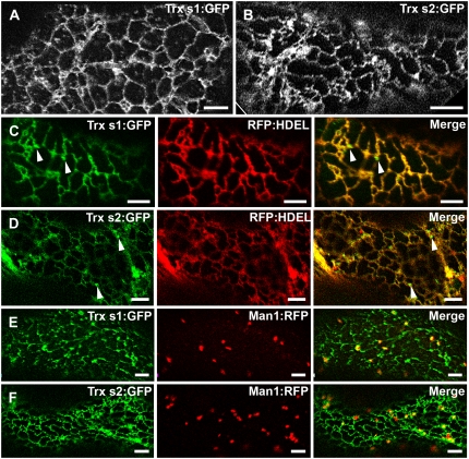
Figure 6.
Visualization of Trxs s:GFP in M. truncatula and N. benthamiana leaf epidermal cells. A, ER-like fluorescence pattern resulting from Trx s1:GFP expression in M. truncatula. B, ER-like fluorescence pattern resulting from Trx s2:GFP expression in N. benthamiana. C and D, Coexpression of Trx s1:GFP and Trx s2:GFP, respectively, with the ER marker RFP:HDEL. In cells coexpressing Trxs s:GFP and RFP:HDEL, near perfect colocalization is observed. Punctuate structures in the ER vicinity are indicated by arrowheads. E and F, Coexpression of Trx s1:GFP and Trx s2:GFP, respectively, with the Golgi marker Man1:RFP. Only very moderate colocalization between Trxs s:GFP and Man1:RFP is observed. C and E, M. truncatula leaf epidermal cells. D and F, N. benthamiana leaf epidermal cells. Scale bars = 5 μm.

