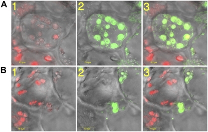
Figure 3.
Citrus Chlase expression versus chlorophyll levels in ethylene-treated lemon fruit monitored by in situ immunofluorescence. Mature green lemon fruit were treated with ethylene (20 μL L−1) at 25°C in the dark for 24 h in a 4-L sealed container. The container was ventilated once after 12 h, followed by injection of fresh ethylene, to maintain CO2 levels below 1%. The flavedo of the treated fruit was dissected and used for obtaining cross sections (70 μm thick) using a vibratome. Tissue sections were fixed and dressed with affinity-purified anti-citrus Chlase antibody (rabbit) followed by Alexa-488-conjugated (goat anti-rabbit) secondary antibody. Fluorescence was visualized using a laser scanning confocal microscope as described in “Materials and Methods.” Images of representative cells (A and B) are presented as follows: 1, confocal image recorded simultaneously in transmitted and red fluorescence mode (i.e. chlorophyll fluorescence superimposed on the bright-field image); 2, confocal image recorded simultaneously in transmitted and green fluorescence mode (i.e. immunofluorescence resulting from the Alexa-488 secondary antibody, corresponding to detection by anti-Chlase antibody, superimposed on the bright-field image); 3, confocal image recorded simultaneously for transmitted, red, and green fluorescence (i.e. red fluorescence corresponds to chlorophyll autofluorescence, green immunofluorescence resulting from the Alexa-488 secondary antibody, corresponding to detection by anti-Chlase antibody, superimposed on the bright-field image). Scale bar = 10 μm (A and B).

