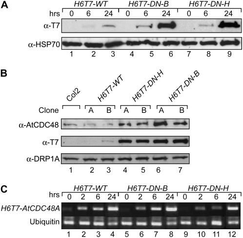
Figure 4.
Expression of conditional dominant-negative Atcdc48A mutants. A, Time course of transgenic ethanol-induced H6T7-Atcdc48 mutant protein expression. Expression of H6T7-AtCDC48A (H6T7-WT; lanes 1–3), H6T7-Atcdc48DN-B (H6T7-DN-B; lanes 4–6), and H6T7-Atcdc48DN-H (H6T7-DN-H; lanes 7–9) proteins was monitored by SDS-PAGE and immunoblot analysis using an anti-T7 antibody (top) and an anti-HSP70 antibody as a protein load control (bottom). Seedling samples were prepared prior to ethanol treatment (lanes 1, 4, and 7) or after 6 h (lanes 2, 5, and 8) or 24 h (lanes 3, 6, and 9) post-ethanol treatment. B, Immunoblot analysis of endogenous AtCDC48 and H6T7-AtCDC48A protein expression levels from untransformed (Col2, lane 1) and independent transgenic ethanol-induced wild-type H6T7-AtCDC48A (H6T7-WT; lanes 2 and 3), H6T7-Atcdc48ADN-H (H6T7-DN-H; lanes 4 and 5), and H6T7-Atcdc48ADN-B (H6T7-DN-B; lanes 6 and 7). Segments correspond to probing of samples with anti-AtCDC48A (top), anti-T7 (middle), and anti-DRP1A (bottom, load control). Samples were processed and analyzed at 48 h postinduction. C, Reverse transcription-PCR analysis of H6T7-AtCDC48A and H6T7-Atcdc48ADN gene expression. cDNA was synthesized from total RNA from H6T7-AtCDC48A (H6T7-WT; lanes 1–4), H6T7-Atcdc48DN-B (H6T7-DN-B; lanes 5–8), and H6T7-Atcdc48DN-H (H6T7-DN-H; lanes 9–12) isolated prior to ethanol treatment (lanes 1, 5, and 9) or after 2 h (lanes 2, 6, and 10), 6 h (lanes 3, 7, and 11), or 24 h (lanes 4, 8, and 12) post-ethanol treatment. cDNA fragments were PCR amplified using primers specific to the transgene (top) or the ubiquitin control (bottom).

