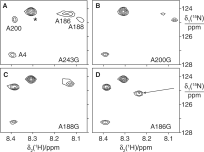
Figure 6.
Assignment of 15N-HSQC spectra of 15N-alanine labeled ɛ:θ complexes by site-directed mutagenesis. ε was labeled with 15N-alanine and θ was unlabeled. The four spectra are of samples prepared with ɛ where different alanine residues were mutated to glycine. (A) A243G mutant. The asterisk identifies a cross-peak that was not observed in the wild-type protein (Figure 3A). It may arise from a low-molecular weight metabolite. (B) A200G mutant. (C) A188G mutant. (D) A186G mutant. The arrow identifies the putative shift of the cross-peak of Ala188 from its position in the wild-type protein.

