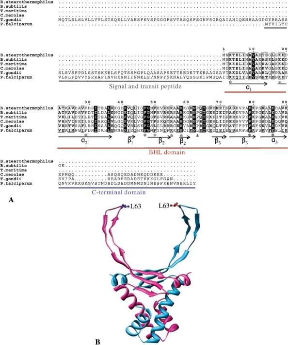
Figure 1.
Sequence alignment and structure model of PfHU. (A) ClustalW alignment of PfHU with bacterial (Bacillus stearothermophilus, B. subtilis, Thermotoga maritima), red algal chloroplast (Cyanidioschyzon merolae) and apicomplexan (Toxoplasma gondii) HU proteins. Conserved residues described in the text are marked with asterisk. (B) Structure of the PfHU dimer modeled on the crystal structure of B. stearothermophilus HU. The position of the leucine residue (L63) that replaces the conserved proline of other HU proteins is indicated. The 42 aa C-terminal domain could not be modeled on any known protein structure and is not depicted in the figure.

