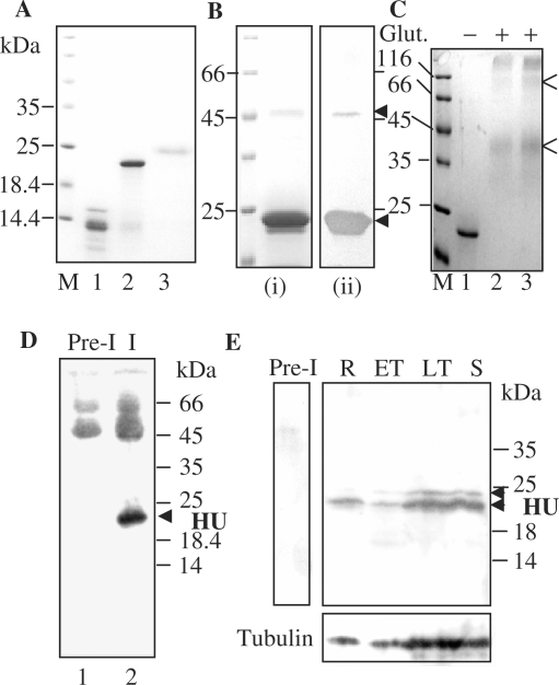
Figure 2.
Expression of recombinant PfHU and its detection in P. falciparum. (A) Purified recombinant proteins PfHUΔC (lane 1), PfHUp (lane 2) and PfHUup (lane 3). M denotes marker lane. (B) Multimeric forms of purified PfHUp seen in Coomassie-stained SDS–PA gels (i) and detected by anti-6XHis antibody in Western blots (ii). (C) Chemical-crosslinking of 5 µg (lane 2) and 7 µg (lane 3) PfHUp indicates dimerization of the protein in solution. Glut., glutaraldehyde. Dimer and tetramer forms are indicated by arrows. (D) Detection of processed HU protein in P. falciparum lysates immunoprecipitated with rabbit anti-PfHUp antibody followed by detection using mouse anti-PfHUp antibody in a Western blot. Lane 1 represents immunoprecipitation with rabbit preimmune serum. (E) Expression of PfHU in P. falciparum intra-erythrocytic stages. The upper and lower panels are Western blots using anti-PfHUp antibody and α, β-tubulin antibodies (Sigma), respectively. R, rings; ET, early trophozoites; LT, late trophozoites; S, schizonts. Pre-I, lysate probed with preimmune serum. Arrows indicate unprocessed and processed forms of PfHU.

