Figure 4.
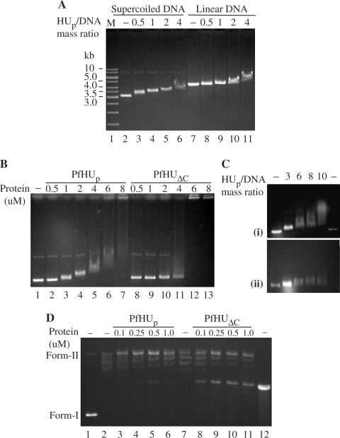
DNA-binding properties of PfHUp. (A) EMSA depicting binding of PfHUp to supercoiled and linear pBR322. Four hundred nanograms of supercoiled (lanes 2–6) or linear (lanes 7–11) plasmid was incubated with PfHUp at different protein/DNA mass ratios. M, DNA marker. (B) EMSA with increasing concentrations of PfHUp (lanes 2–7) and PfHUΔC (lanes 8–13) using supercoiled pBR322 DNA. Lane 1 is free DNA. (C) Binding of PfHUp to supercoiled DNA in the presence of 50 mM (i) and 100 mM (ii) NaCl. (D) DNA supercoiling assay with increasing concentrations of PfHUp (lanes 3–7) and PfHUΔC (lanes 8–12). Lane 1 is naked DNA (negatively supercoiled pBR322), lane 2 is pBR322 partially relaxed with topoisomerase I, lane 12 is linearized pBR322. Form-I, supercoiled DNA; form-II, relaxed DNA.

