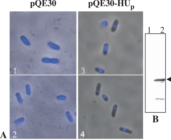
Figure 5.

PfHUp-mediated condensation of E. coli nucleoids. (A) Fluorescence images of DAPI-stained, IPTG-induced E. coli cells that had been co-transformed with pQE30 + RIG plasmid (1 and 2) or pQE30-HUp + RIG plasmid (3 and 4). (B) Western blot using anti-PfHUp antibody to detect expression of PfHUp in E. coli cells co-transformed with RIG and pQE30 (lane 1) or pQE30-HUp (lane 2) followed by induction with IPTG. Arrow indicates the PfHUp band.
