
Fig. 2.
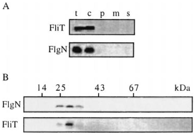
Characterization of the cellular pools of FlgN and FliT.
A. Location. Late exponential phase cultures of WT S. typhimurium were harvested, and cells were fractionated. Proteins from total cells (t), culture supernatant (s) and the cytosolic (c), periplasmic (p) and membrane (m) fractions were separated by SDS–(15%)PAGE and immunoblotted with anti-FlgN or anti-FliT antisera.
B. Size. Cytoplasmic cell extracts were loaded onto a calibrated Superose 12 10/30 HR column and developed by FPLC. Eluted fractions were immunoblotted with anti-FlgN or anti-FliT antisera. Peak fractions of eluting size markers are indicated (BSA, 67 kDa; ovalbumin, 43 kDa; chymotrypsinogen, 25 kDa; ribonuclease A, 14 kDa).